September 10, 2012

Biparietal foramen

Biparietal foramen are not an uncommon finding on a routine skull radiograph, but occasionally they can become "giant," simulating a lytic lesion.

Giant biparietal neuroforamen: The most important thing about this variant is not to mistake it for disease.  These foramina are not uncommonly detected in children, and a high-radiation dose CT can be avoided if the variant is recognized.


According to available sources, these foramina are heritable in an autosomal dominant pattern, and represent part of a spectrum of bifid cranium (bifid cranium at one extreme and parietal foramen at the other).

------------------

1. Fein JM, Brinker RA. "Evolution and significance of giant parietal foramina" J.  Neurosurg. Vol:37. October, 1972.
2. Little BB, Knoll KA, Klein VR, et al. "Hereditary Cranium Bifidum and Symmetric Parietal Foramina Are the Same Entity." American Journal of Medical Genetics 35:453-458 (1990)

May 23, 2012

Greyhound skull


Skaphe (boat) + cephalus (head)  
&  Dolikhos (long) + cephalus (head)

Scaphocephaly/Dolichocephaly are descriptive terms for a skull that is abnormally longer than it is wide, with a cephalic index <75 for women and < 75.9 for men. Scaphocephaly/Dolichocephaly is the most common kind of simple craniosynostosis, comprising 40-55% of nonsyndromic cases.  Craniosynostoses occur with a frequency of 1:2000-2500.

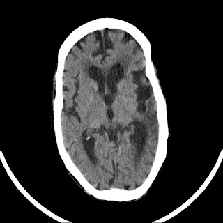
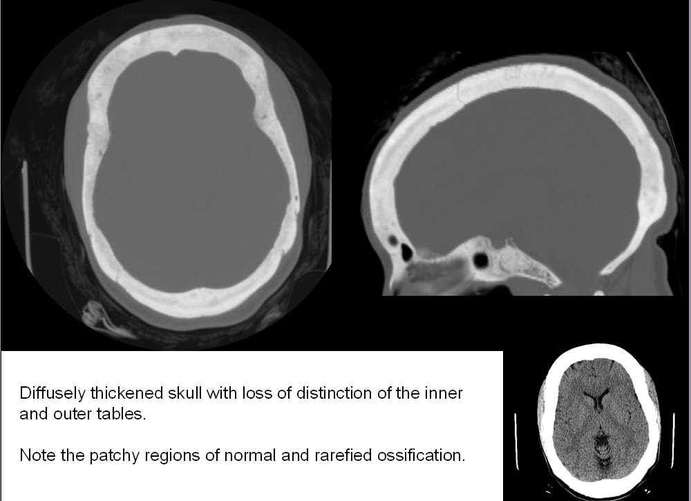
Scaphocephalic/Dolichocephalic skull
(old left MCA infarct as well)
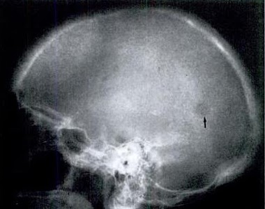
Scout radiograph of same patient

The cephalic index was originally devised by Anders Retzius (the same with the "space" named after him).  It's simply a ratio of the widest AP and TV distances of the skull.  (The TV distance is the same as the biparietal diameter, BPD). Virchow (the same with the "triad" named after him) pointed out that when a suture closes prematurely (in scaphocephaly, the sagittal), then the growth of the skull is restricted in the plane perpendicular to the suture, and compensate in the parallel direction.

The scaphocephaly demonstrated above is a simple form of craniosynostosis... simple in the sense that only one suture has prematurely fused, the sagittal.  Other simple craniosynostosis include brachycephaly (premature fusion of the coronal suture) and trigonocephaly ("keel skull," premature fusion of the metopic suture). Compound craniosynostosis involves more than one suture, such as in plagiocephaly ("skew skull").

Although certainly a morphologic/aesthetic issue, and frequently related to congenital syndromes, the craniosynostoses may also occur by themselves and have no other associated effects. One source mentions that the formation of craniosynostoses may have an effect on the underlying CSF spaces -- decreased/eliminated at the premature suture line, excessive in the areas of abnormally increased skull space

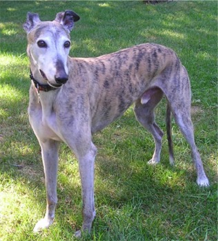
The concept of "appropriate" skull size is species-specific, since some animals are supposed to have skulls longer than they are wide... and you could just as easily call scaphocephaly "greyhound skull" since the greyhound is one of the many dogs whose heads are normally scaphocephalic:

The greyhound: a normally scaphocephalic dog


Although the two terms scaphocephaly and dolichocephaly are synonymous, the term dolichocephaly may be a more consistent fit with the other end of the spectrum, brachycephaly ("short skull").  Incidentally, between the two lies the more or less useless term mesaticephaly.

-----

1. Benson ML, Oliverio PJ, Yue NC, Zinreich SJ. Primary Craniosynostosis: Imaging Features. AJR 1996;166:697-703 036
2. Delashaw JB, et al. Cranial Vault Growth in Synostosis: Review Article. JNS  70:159-165, 1989

November 22, 2011

The Vidian Canal

The vidian canal is a landmark in the skull base that transmits the vidian nerve and vidian artery.


The vidian nerve is formed from the greater/large superficial petrosal nerve of CN V (sympatheticsand the deep petrosal branch (parasympathetics).   (There's also an ascending sphenoid branch from the otic ganglion).


The vidian canal transmits the nerve from the foramen lacerum to its exit at the pterygopalatine fossa.


Tracing the vidian canal superior to inferior.  It originates at the pterygopalatine fossa (arrow) and proceeds through the skull base (circle).  The branch point in the third image represents the take-off of the GSPN into the middle ear through the "hiatus" of the greater "superficial" petrosal nerve (longer left branch).  The smaller branch represents the deep petrosal nerve joining the GSPN with its post synaptic carotid nerve fibers.

Three variations of the vidian canal have been described with varying degrees of bony coverage (below).


Incidentally, The term vidian is derived from an anatomist, "Vidus."


November 5, 2011

Renal osteodystrophy and diffuse skull thickening

Although not difficult to detect, the challenge in diffuse skull thickening is suggesting a correct etiology from a wide range of possibilities.


The range of possible etiologies includes:
- Normal variant (most common etiology), Acromegaly, Microcephaly
- Chronic shunting
- Chronic Dilantin/Phenytoin use
- Diffuse osteoblastic metastases
- Paget disease
- Extramedullary hematopoeisis (esp. B-thalassemia)
- Osteopetrosis
- Hyperparathyroidism (either primary or secondary)

In the example above, the patient had a known diagnosis of renal osteodystrophy, and the thickened calvaria were attributed to that cause.

Findings in diffuse skull thickening from renal osteodystrophy are described as "loss of distinction between the inner and outer table" and a "granular deossification of the skull" ("salt and pepper skull") due to small lytic foci interspersed with normal bone. (below)


Brown tumors can occasionally occur in this setting (below).  Cranial neuropathy from narrowing of foramina has also been described.

"Salt and Pepper" appearance of the skull with a focus of brown tumor (arrow).

------
1. Baird IM,  Lees F, "Renal Osteodystrophy in Adults" AMA Arch Intern Med. 1956;98(1):16-22. 
2. Burgener F, et al. "Differential Diagnosis in Conventional Radiology" 3rd ed (2008), pp. 218-219

October 30, 2011

The Double Transverse Foramen

The double transverse foramen in the cervical spine is a normal variant that, according to the literature, occurs most often in the C6 vertebra transverse process, with decreasing frequency above and below.  It is claimed that the vertebral artery splits at these accessory foramen (which are always smaller), and rejoins distally, although this is controversial, and it is not entirely clear from the literature whether the accessory foramen usually contains artery, vein, nerve, neither, or some combination. 

Bilateral transverse foramina incidentally noted at C5 on this trauma patient.
Bilateral transverse foramina also incidentally noted at C6 on this trauma patient.  C6 is the most common level to find double transverse foramina.

One source claims that accessory foramina occur in 1.6% of observed cervical spines. The duplication is thought to represent the failure of controlled regression of two intersegmental arteries and a segment of the primitive dorsal aorta.  An extremely rare triple transverse foramen has been reported.


1. Francis CC. "Dimensions of the cervical vertebrae" The Anatomical Record. Anat. Rec. Vol 122: 4 pp.  1097-0185
2. Wysocki J, Bubrowski M, Reymond J, Kwiatkowski J. "Anatomical variants of the cervical vertebrae
and the first thoracic vertebra in man" Folia Morphol. Vol. 62, No. 4, pp. 357–363
3. Murlimanju BV, et al. "Accessory Transverse Foramina in the Cervical Spine: Incidence, Embryological Basis, Morphology and Surgical Importance" Turkish Neurosurgery 2011, Vol: 21, No: 3, 384-387

October 22, 2011

Luckenschadel

Lucke (gap) + schadel (skull)

The luckenschadel (sometimes called "luckenschadel skull," although this is technically redundant) is a calvarial abnormality most often associated with a Chiari II malformation and a meningomyelocele

The luckenschadel is an ossification disorder which leads to multiple deep pits in the membranous bones of the newborn skull (favoring the parietal and occipital bones).  It has a classic radiographic "honeycomb" appearance (below), which is diagnostic. 

Alternating regions of cortical fenestration, creating a "honeycomb" appearance, compatible with Luckenschadel.  There is also scalp swelling.  Vaginal delivery of these infants can be problematic since cranial pressure on a fenestrated skull can lead to intracranial hemorrhage.
3D CT reformats of luckenschadel

The grooves in the skull do not necessary traverse the entire bone, and when limited to the inner table, they are called "craniolacunae."  If palpable and involving both the inner and outer table, they are then referred to as "craniofenestrae."  The "waviness" of the skull outline on axial imaging is detectable on obstetric ultrasound (below).


Developmentally, luckenschadel is similar to myelomeningocele, in that there is failure of normal bone formation over the brain.  Some sources claim that it develops in part due to lack of normal cerebral ventricular distention due to an open neural tube defect in the Chiari II malformation.  Over the first 6 months, the fenestrae fill in, and luckenschadel is a diagnosis that can only be made in neonates.


1.   Vigliani, M.  "The luckenschadel skull: a forgotten entity"  Obstetrics and Gynecology: Volume 111(2, Part 2), February 2008, pp 562-565
2.  Coley BD. "Ultrasound diagnosis of luckenschadel (lacunar skull)" Pediatric Radiology, Volume 30:2. pp 82-84.

October 17, 2011

The "Two-eyed Scotty Dog"


The vertebral transverse process contains two additional processes of its own.  The more superior (and larger) of the two is called the mammillary process, which connects posteriorly to the superior articular facet, and is in the same plane as the transverse process.


There is normal variation in the size of this process, but occasionally, a large mammillary process in a sufficiently obliqued radiograph will give the appearance of the "two-eyed scotty dog," with one eye being the usual pedicle and the other eye being the mammillary process.

Oblique view showing the "two-eyed Scotty dog" appearance at L3.  The anterior ring-shaped opacity represent the pedicle (large arrow); the more posterior ring-shaped opacity represents a prominent mammillary process (small arrow).

1.  Patel NP, Kumar R, Kinkhabwala M, Wengrover S. "Radiology of lumbar vertebral pedicles: variants, anomalies, and pathologic conditions"  Radiographics. Vol 7:1 (Jan 1987) pp. 101-137.